The seventh edition of this classic work makes mastering large amounts of information on the nervous system and sensory organs much easier. It provides readers with an excellent review of the human body and its structure, and it is an ideal study companion as well as a thorough basic reference text. The many user-friendly features of this atlas include: New and enhanced clinical tips Hundreds of outstanding full-color illustrations with updated labels Side-by-side images with explanatory text Helpful color-coding and consistent formatting throughout Emphasizing clinical anatomy, this atlas integrates current information from a wide range of medical disciplines into discussions of the nervous system and sensory organs, including: In-depth coverage of key topics such as molecular signaling, the interplay between ion channels and transmitters, imaging techniques (e.g., PET, CT, and NMR), and much more A section on topical neurologic evaluation Volume 3: Nervous System and Sensory Organs and its companions Volume 1: Locomotor System and Volume 2: Internal Organs comprise a must-have resource for students of medicine, dentistry, and all allied health fields.
W. Werner Kahle Libros
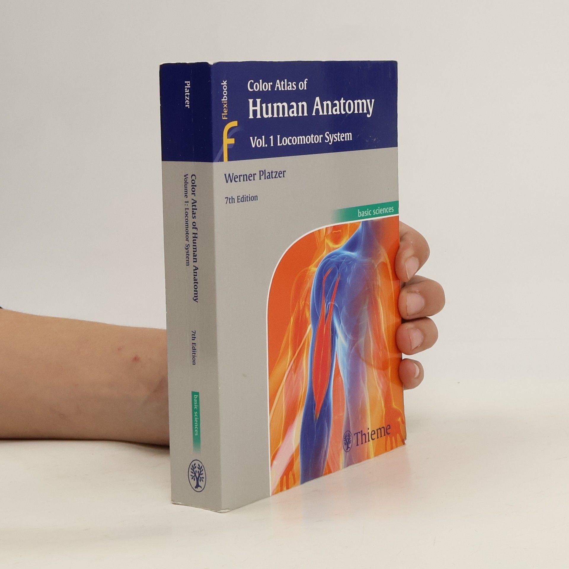

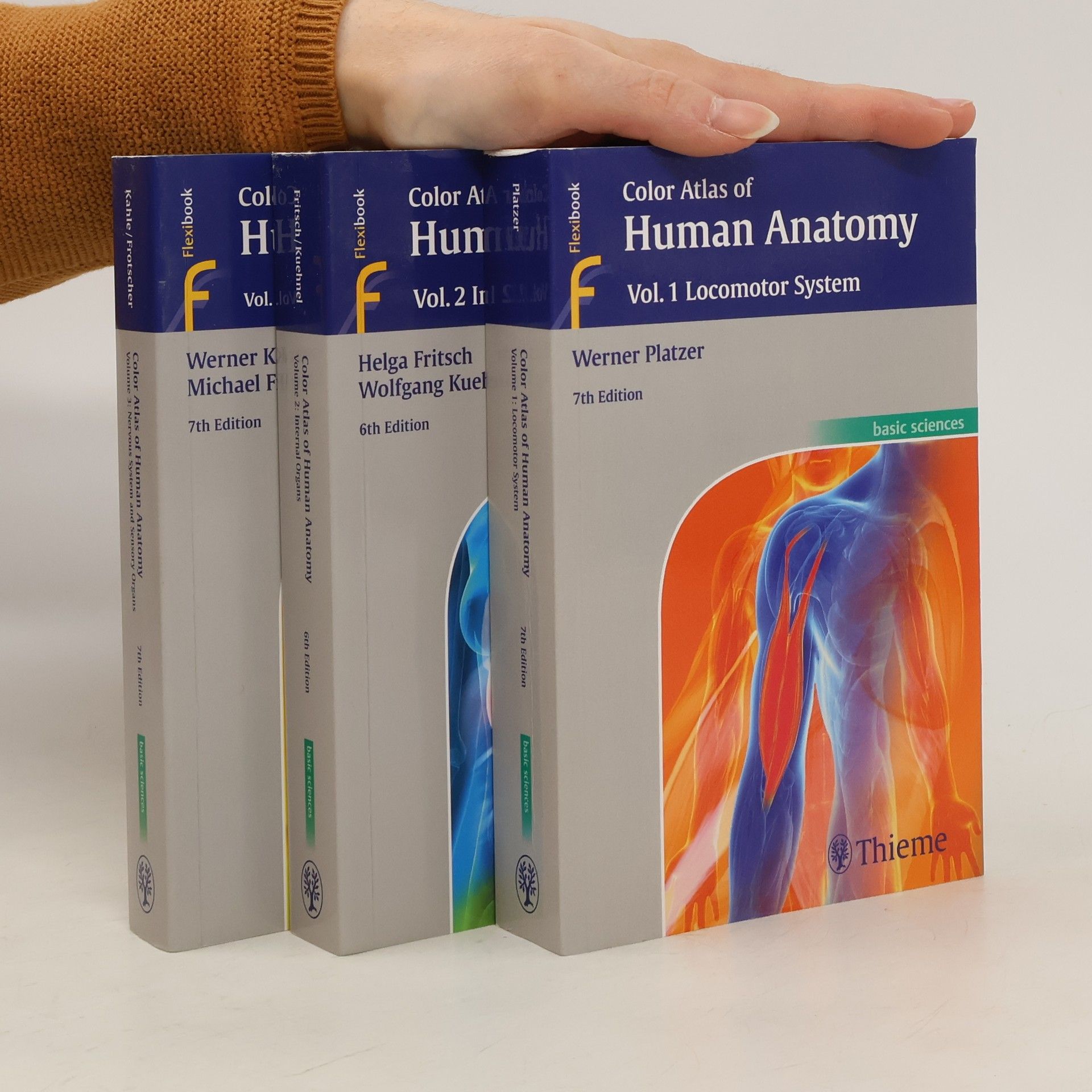
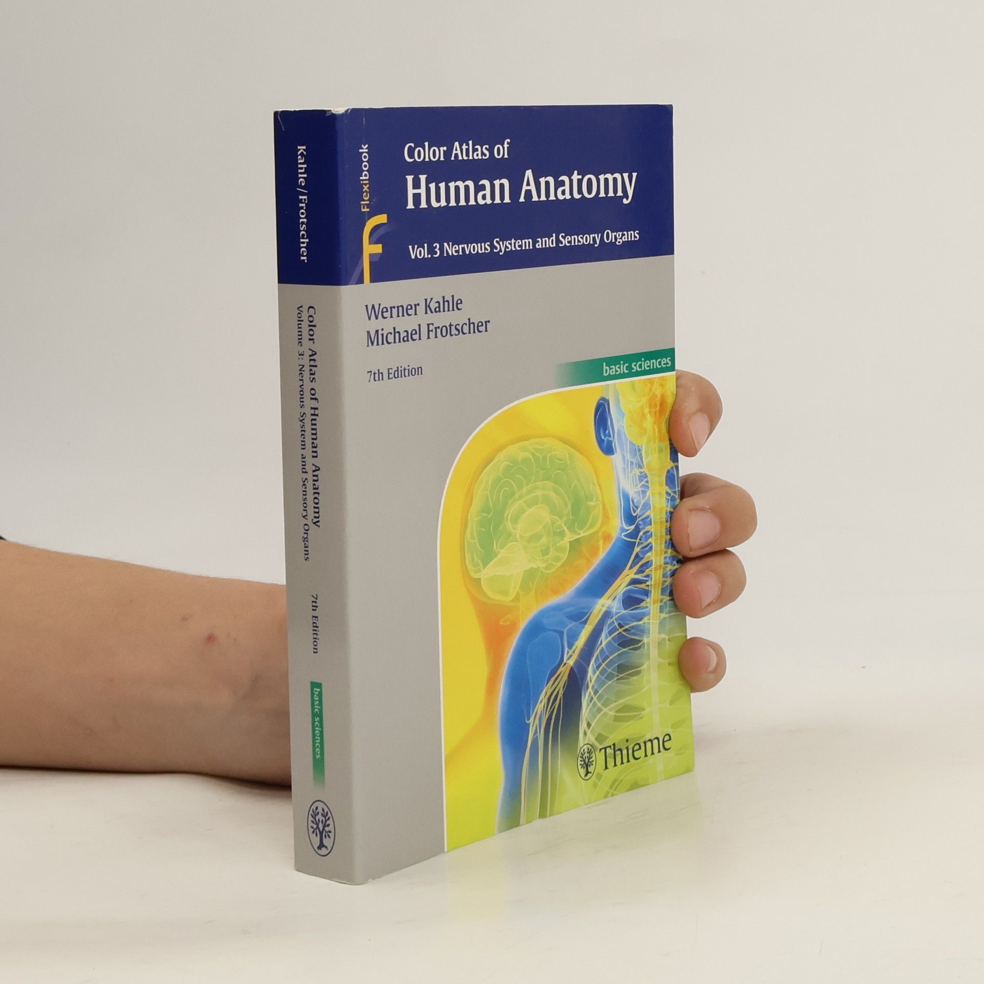
Color Atlas of Human Anatomy 1 - 3
Locomotor System. Nervous System and Sensory Organs. Internal Organs
- 3 volúmenes
Color Atlas of Human Anatomy, Volume 3: Nervous System and Sensory Organs For over 45 years, the three-volume Color Atlas of Human Anatomy has provided readers with a compact review of the human body and its structures. It is ideal for studying, preparing for exams, and as a reference. The new, 8th edition of Volume 3: Nervous System and Sensory Organs builds on a robust foundation of scientific knowledge, summarizing in its compactness the structure and functions of the nervous system and sensory organs. Key highlights: Updated to include the latest findings in neuroanatomy Proven concept of concise texts paired with 190 color plates of outstanding anatomical illustrations The structure and topography of the various components of the nervous system and their complex, functional interactions are explained Important neuroanatomical research techniques and the use of imaging methods (CT, MRI, PET, and SPECT) are discussed Volume 3: Nervous System and Sensory Organs is accompanied by Volume 1: Locomotor System (ISBN 978-3-13-242443-3) and Volume 2: Internal Organs (ISBN 978-3-13-242448-7).
A sound understanding of the structure and function of the human body in all of its intricacies is the foundation of a complete medical education. This classic work makes the task of mastering this vast body of information easier and less daunting with its many user-friendly features: Hundreds of outstanding full-color illustrations; Clear organization according to anatomical system; Abundant clinical tips; Side-by-side images and explanatory text; Helpful color-coding and consistent formatting throughout; and Useful references and suggestions for further reading. Emphasizing clinical anatomy, the text integrates current information from an array of medical disciplines into the discussions of the nervous system and sensory organs, including: In-depth coverage of key topics, including molecular signaling, the interplay between ion channels and transmitters, imaging techniques such as PET, CT, and NMR, and much more with a full updated section on topical neurologic evaluation. -- Publisher
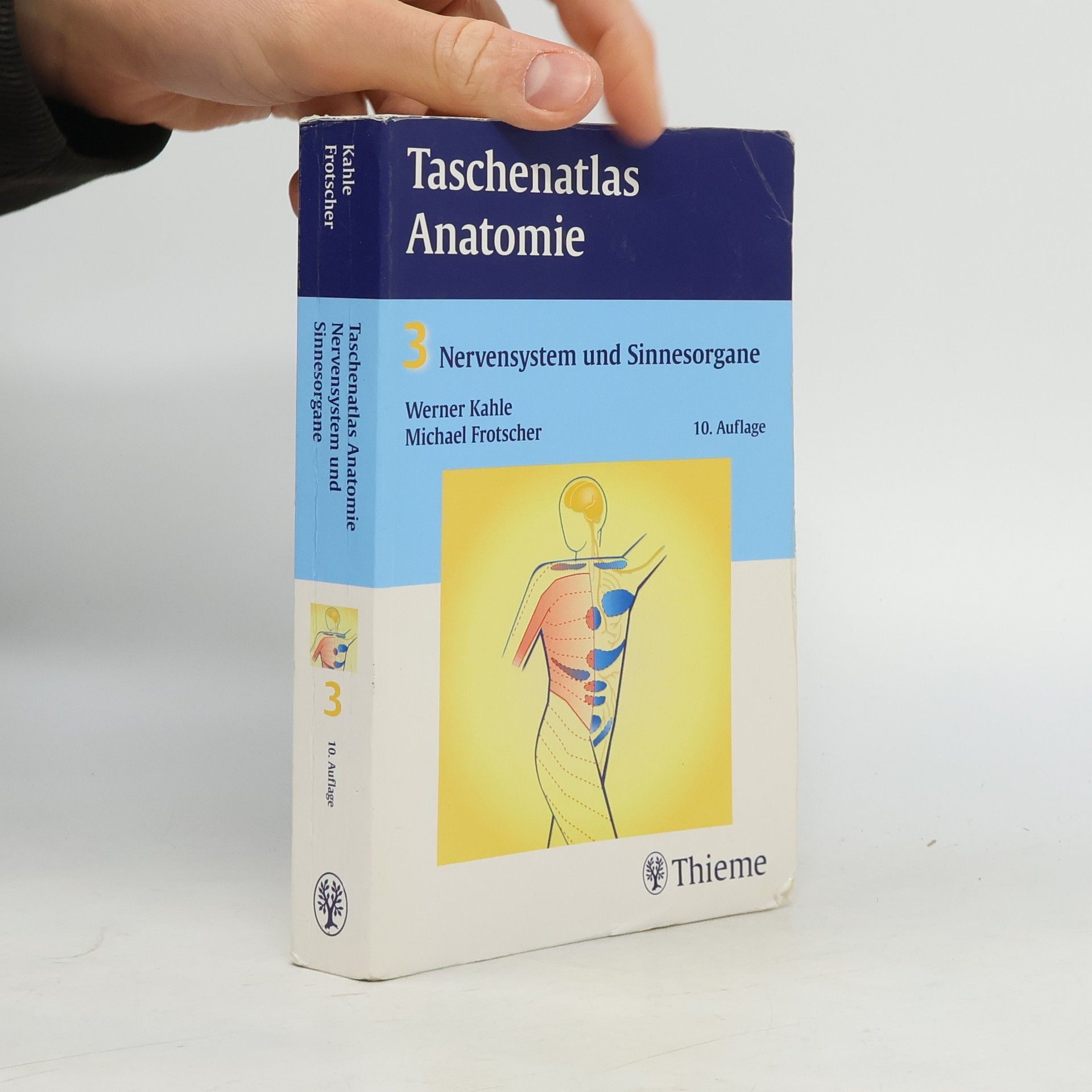
Taschenatlas Anatomie 03. Nervensystem und Sinnesorgane - 10. Auflage
- 423 páginas
- 15 horas de lectura
Anatomie in Wort und Bild Dieser 3-bändige Taschenatlas bietet Ihnen einen anschaulichen Überblick über den Aufbau des menschlichen Körpers. Das bewährte Konzept aus Textseite und Bildtafel ist ideal zum Lernen, zur Prüfungsvorbereitung und zum Nachschlagen geeignet. Viele klinische Bezüge im Text vermitteln Ihnen den Bezug zur Praxis. Band 3: Nervensystem und Sinnesorgane - Dieser Band gibt Ihnen einen systematischen Überblick über den Aufbau und die funktionelle Organisation des Nervensystems. - Wichtige Techniken der anatomischen Forschung und bildgebende Verfahren (PET, CT, NMR) werden kompakt dargestellt, ebenso die molekularen Grundlagen der Erregungsübertragung und das Zusammenspiel von Ionenkanälen und Transmittern. - Häufig verwendete Synonyme werden im Text und im Sachverzeichnis zusammen genannt, so dass Sie die vielen bedeutungsgleichen Begriffe leicht erlernen können. Kompakt und komplett - Ihr verlässlicher Begleiter durch die Anatomie!
Dtv-Atlas der Anatomie 3. Nervensystem und Sinnesorgane
- 381 páginas
- 14 horas de lectura
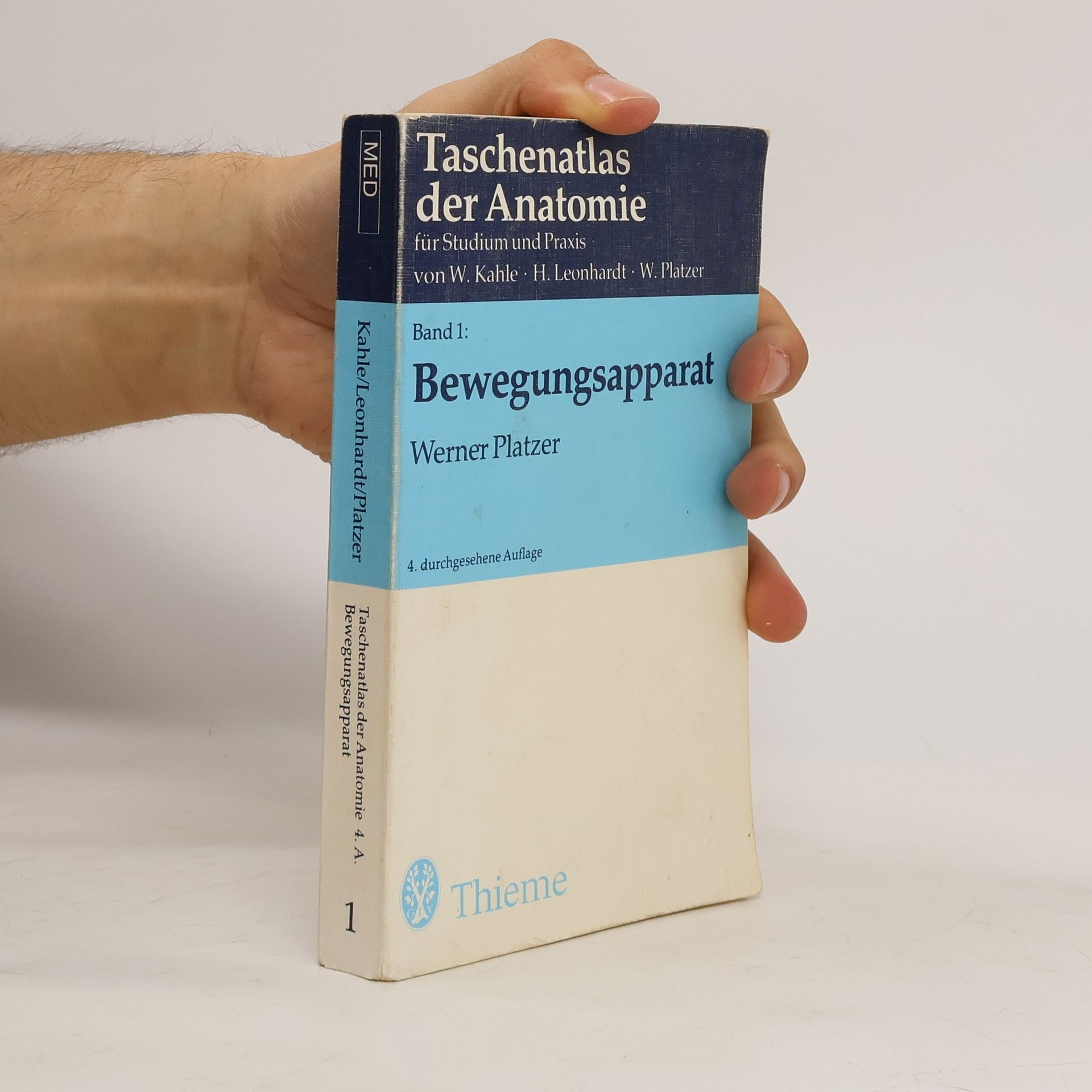
Taschenatlas der Anatomie für Studium und Praxis 1
Bewegungsapparat
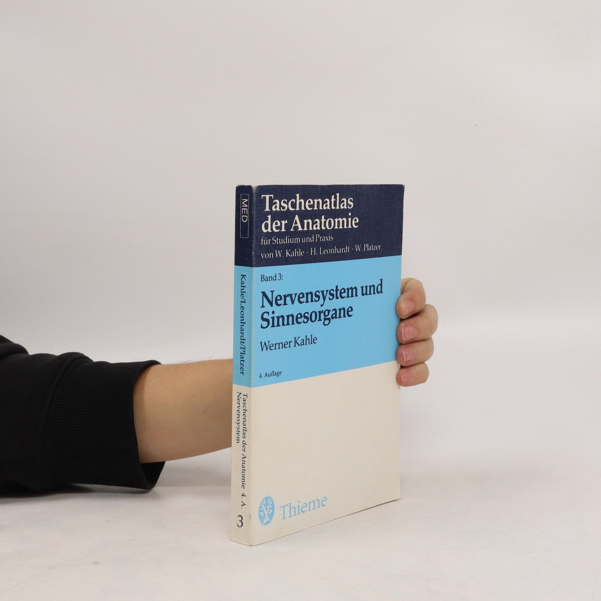
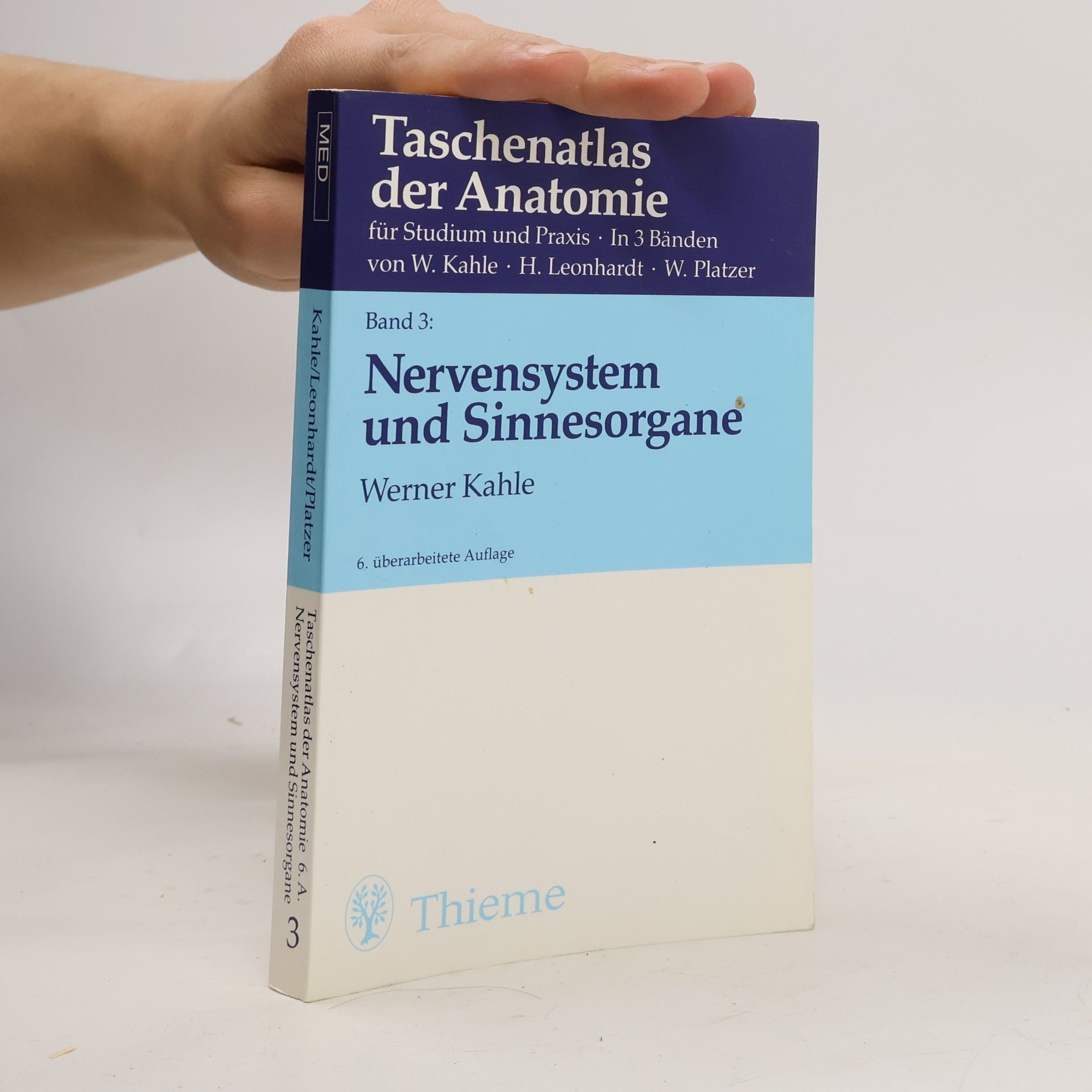
Taschenatlas der Anatomie für Studium und Praxis
Band 3. Nervensystem und Sinnesorgane
